UVP iBox in vivo BioImaging Systems
Analytik Jena
Easily in vivo small animal imaging system enables research studies in whole animal down to individual cells

Systems enables non-invasive detection of fluorescent and bioluminescent reporters in small animals. Analytik Jena US offers three instrument series:
- UVP iBox Studio
- UVP iBox Scientia
- UVP iBox Explorer2
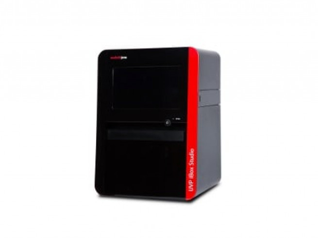
1. UVP iBox Studio
- Affordable small animal imaging
- Integrated 13.3” touch screen computer
- Non-invasive detection of fluorescent reporters in up to 3 mice
- High resolution, high sensitivity, deeply cooled CCD cameras (up to -60°C from ambient)
- iBox Studio 815: 8MPx camera, good sensitivity in the whole spectrum
- iBox Studio 695: 6MPx camera, highly effective in NIR
- Wide aperture (f/0.95) lens optics capture more light in low-light applications
- Overhead white, green, red and blue LEDs come as standard in the series
- Optional NIR and eLITE multispectral light source
- 5-Position automated filter wheel (GFP and RFP emission filters included)
- Small footprint to maximize the use of laboratory bench space
- Light tight darkroom creates optimum imaging conditions
- Warming plate
- Roll-out tray provides easy access for placement of mice on the warming plate
- Optional anesthesia system
- VisionWorks Software, with comprehensive features, optimizes image acquisition and analysis
- Automated templates for repeated experiments
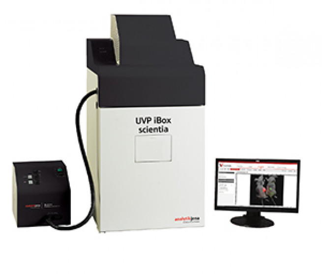
2. UVP iBox Scientia
- Small Animal Imaging System
- Non-invasive detection of fluorescent and bioluminescent reporters in up to 5 mice
- Selection of high resolution, high sensitivity CCD cameras for in vivo imaging
- BioCam 900: 1.0Mpx, quantum efficiency >90 %, Back-illuminated chip, cooled -70°C from RT
- OptiChemi 695: 6.0 Mpx, high efficiency and sensitivity, especially in NIR, cooled -60°C from RT
- Wide aperture (f/0.95) lens optics capture more light in low-light applications
- Illumination: overhead white LED and UV, eLITE multispectral light source
- 5-Position automated filter wheel
- Matched emission/excitation filter sets specifically for small animal imaging (GFP and RFP included)
- Light tight darkroom creates optimum imaging conditions
- Unique viewing window enables quick sample inspection
- Warming plate
- Roll-out tray provides easy access for placement of mice on the warming plate
- Optional anesthesia system
- VisionWorks Software for analysis and image composition
- Automated templates for repeated experiments
Applications
- Tumor studies
- Cancer research
- Heart disease
- Gene therapy
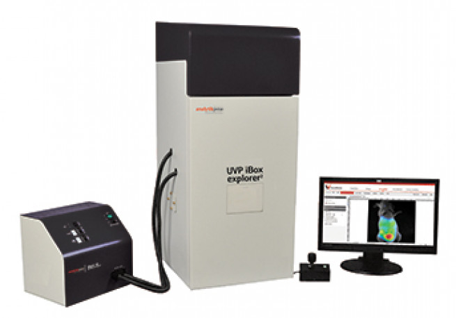
3. UVP iBox Explorer2 Imaging Microscope
- Easily detects GFP/RFP and other fluorescent markers in small animals
- View macro to micro in the whole animal to individual cell
- Able to image targets subcutaneously and within the body cavity of mice
- Motorized stage position with the joystick to adjust positioning at the X, Y, Z-axis
- The upright optics provide an ultra long working distance
- Magnification ranges of 0.17x - 16.5x enables transition from the macroscopic to the microscope scale
- High numerical aperture (NA) for detailed fluorescent in vivo imaging
- OptiChemi 695: 6.0 Mpx, cooled -60°C from RT, high frame rate CCD camera
- Quick detection, image capture and high throughput
- Bright illumination of samples produces an intense fluorescent signal and fast exposure times
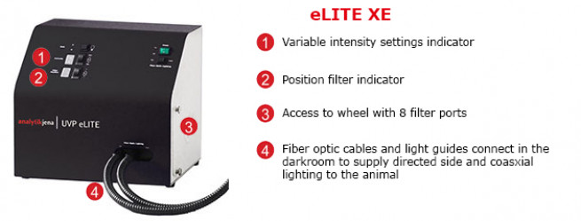
- Easy installation of the UVP eLITE Xe external light source through the darkroom access ports
- Epi fiber optic Xenon light source supplies uniform and directed lighting to the animal
- GFP and RFP excitation filters included
- 4-Position emission filter rack: GFP, RFP, 515 nm Longpass, and Neutral Density filters included
- VisionWorks Software for image composition, capturing and accurate data generation
Applications
- Tumor shedding
- Tumor/host margins and interactions
- Hematogenous trafficking
- Tumor angiogenesis
- Tumor micro environment
- Intralymphatic trafficking
- Micro/macro metastases
- Primary tumor growth
- Biodistribution
Documents and resources
For more information about the UVP iBox Studio Affordable Small Animal Imaging visit producer's website.
For more information about the UVP iBox Scientia Small Animal Imaging System visit producer's website.
For more information about the UVP iBox Explorer2 Imaging Microscope visit producer's website.
Related products

UVP GelStudio Series
Analytik JenaDocumentation system with high sensitivity imaging for wide range genomic and proteomic applications
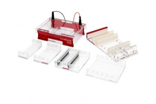
Electrophoresis Techniques
Analytik JenaComprehensive range of instrumentation for polyacrylamide and agarose gel electrophoresis

BlotCycler - Automated Western Blotting
High quality consistent western blot processing saves time and improves your western blotting
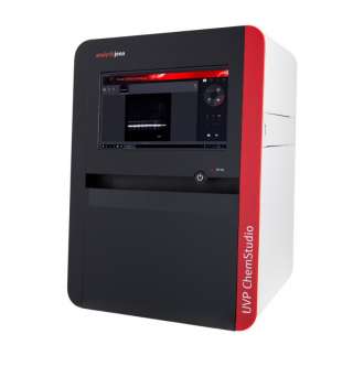
UVP ChemStudio Series
Analytik JenaNext generation imagers for chemiluminescence, fluorescence and colorimetry genomic and proteomic applications